Topographic sketch. Coronal view of bilateral DRT (orange). Patient
Por um escritor misterioso
Descrição

PDF) Three-dimensional LASIK flap thickness variability: Topographic central, paracentral and peripheral assessment, in flaps created by a mechanical microkeratome (M2) and two different femtosecond lasers (FS60 and FS200)

Root Canal Configuration - GRIN

Journal of Comparative Neurology, Systems Neuroscience Journal

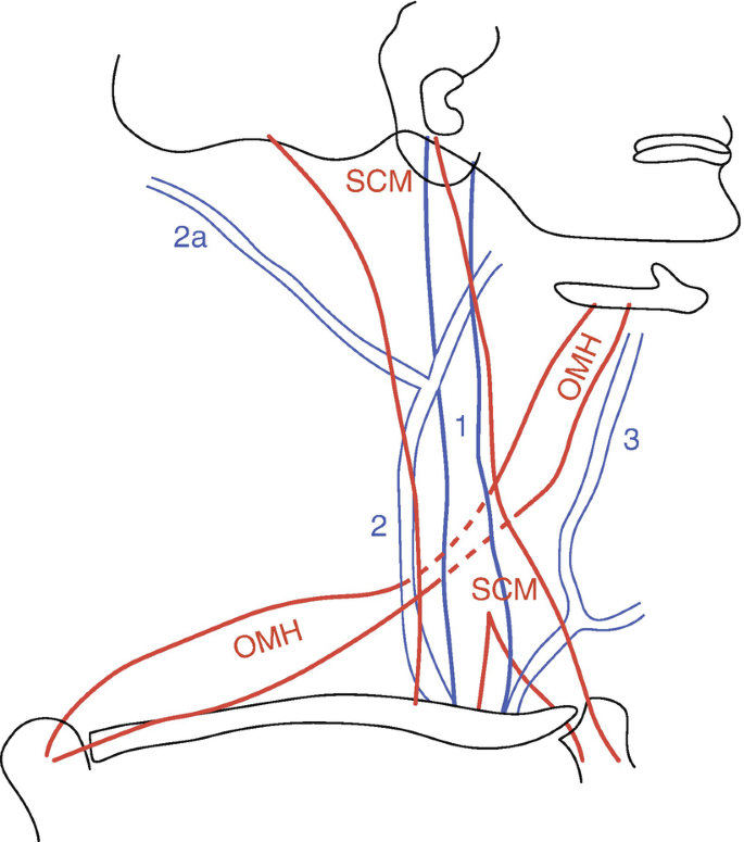
Crossing nerve transfer drives sensory input–dependent plasticity for motor recovery after brain injury
Sensors April-1 2023 - Browse Articles
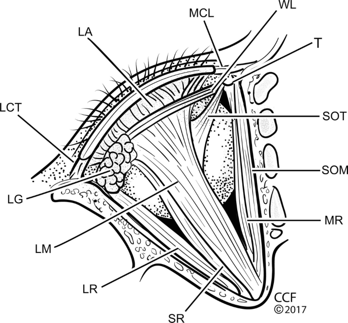
Applied Surgical Anatomy of the Ocular Adnexa
Diagnosis and Differential Diagnosis of Voice Disorders

Advances in human intracranial electroencephalography research, guidelines and good practices - ScienceDirect

ImagineNano2018 Conference Abstracts Book by Phantoms Foundation - Issuu

DTI for brain targeting: Diffusion weighted imaging fiber tractography—Assisted deep brain stimulation - ScienceDirect
de
por adulto (o preço varia de acordo com o tamanho do grupo)