Figure 5 from Retrognathic maxilla in Habsburg jaw
Por um escritor misterioso
Descrição
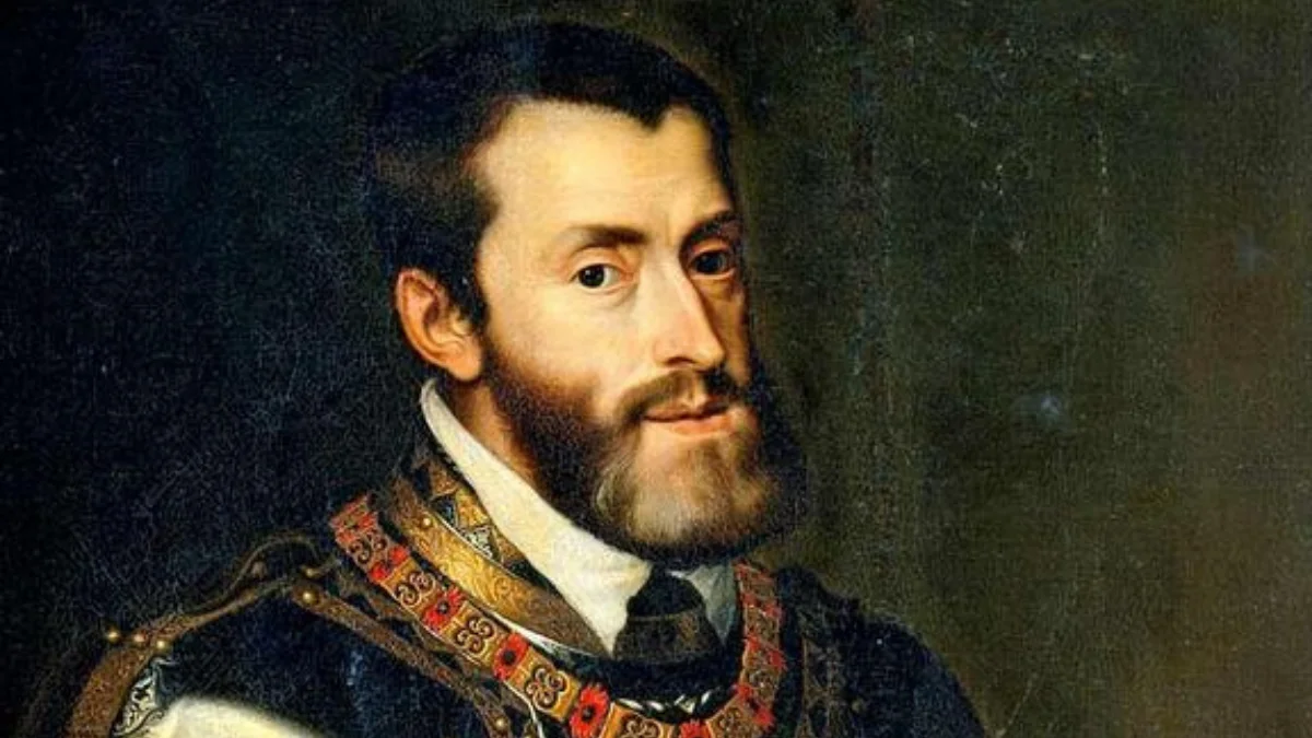
Figure 5. Portrait of Joanna of Austria by Alessandro Allori, 1570, Museo degli Argenti, Firenze, Italy. Image used with the permission of the Soprintendenza Speciale al Polo Museale Fiorentino, Firenze, Italy – Medici Project. - "Retrognathic maxilla in "Habsburg jaw". Skeletofacial analysis of Joanna of Austria (1547-1578)."

a) Retrognathic maxilla of 4 mm; b) Prognathic maxilla of 2 mm; c)

a) Lateral radiograph of the skull (reversed gray scale) obtained of
16 Class III Malocclusion— The Evidence on Diagnosis and Treatment

Prognathism - Wikiwand

Improving Esthetic and Functional Outcomes of Severe Habsburg Jaw Using Modified Mandibular C-Osteotomies: A Case Report and Review of Literature - Andrew M. Henry, Jason W. Yu, Brian B. Farrell, 2021
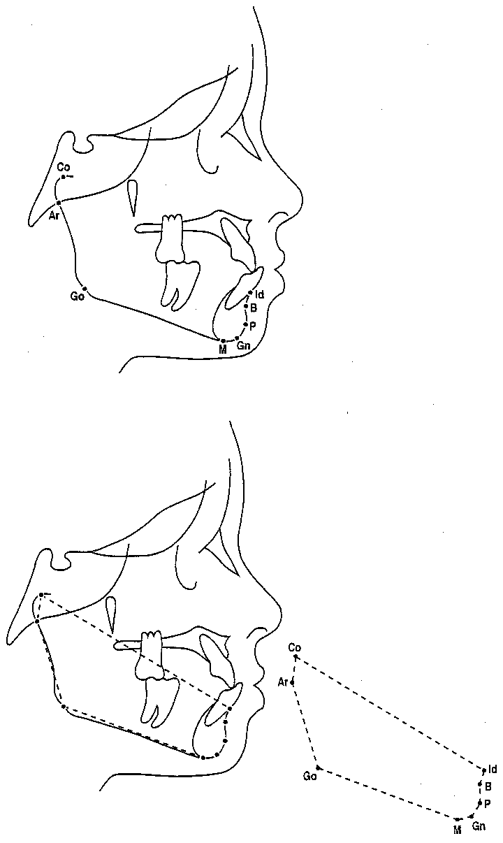
Mandibular landmarks superimposed on a lateral cephalographic tracing

Figure 5 from Retrognathic maxilla in Habsburg jaw. Skeletofacial analysis of Joanna of Austria (1547-1578).

Figure 3 from Cephalometric investigation of Class III dentoalveolar malocclusion.

a) Lateral radiograph of the skull (reversed gray scale) obtained of

Cephalometric investigation of Class III dentoalveolar malocclusion.
de
por adulto (o preço varia de acordo com o tamanho do grupo)